
Ex/Em:328/533 nm
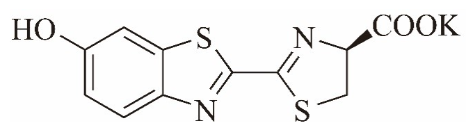
CAS:115144-35-9
分子式:C11H7N2O3S2K
分子量:318.4
1. 基本性質與原理
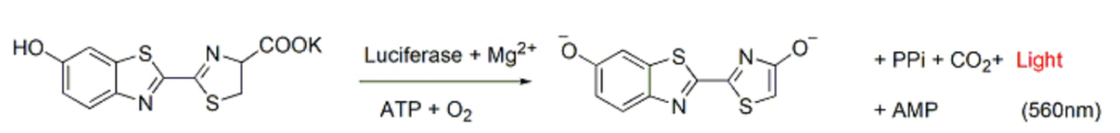
D-Luciferin 是一種自然界存在的化合物(最早提取自螢火蟲)。在科學實驗中,為了增加其溶解度與穩定性,通常會製成**鉀鹽(Potassium Salt)**或鈉鹽形式。
- 發光原理: 在 ATP 和鎂離子(Mg+2)存在的環境下,**螢光素酶(Luciferase)**會催化 D-Luciferin 氧化。這個生化反應會產生激發態的氧化螢光素(Oxyluciferin),當它回到基態時,會釋放出波長約 560nm(黃綠光) 的光子。
- 體內偏移: 雖然在體外反應是黃綠光,但在哺乳動物體內,由於組織吸收與散射,觀測到的發光峰值通常會偏移至 600nm 以上(紅光/近紅外區)。這類長波長光線對組織的穿透力較強,這也是它適合用於活體成像的主因。
2. 為什麼選擇 SYD「D-Luciferin Potassium Salt」?
- 高溶解度: 鉀鹽在水或緩衝溶液(如 PBS)中溶解度極佳,方便配製高濃度儲備液。
- 生物相容性: 對於活體動物(如小鼠、大鼠)的毒性極低,適合腹腔注射(i.p.)或靜脈注射(i.v.)。
- 高純度要求: 活體成像對底物純度要求極高(通常需 >99%),否則雜質可能干擾信號或引起細胞毒性,SYD 生產步驟特別移除去氫螢光素(dehydroluciferin)和 L-螢光素 (L-luciferin)等螢光素酶的競爭性抑制劑。L-luciferin 會抑制訊號強度。
References:
1. “D-luciferin potassium salt (catalogue #MB102, Syd Labs Inc., Natick, MA) was resuspended in phosphate-buffered saline (PBS) to a final concentration of 20 mg/ml. Mice were dosed with 200 μL luciferin substrate intraperitoneally and waited for 10 minutes to allow for distribution of substrate prior to image acquisition.” Interleukin-17 promotes metastasis in an immunocompetent orthotopic mouse model of prostate cancer. David Cunningham, Qiuyang Zhang, Sen Liu, Keshab R Parajuli, Qiang Nie, Lin Ma, Allen Zhang, Zhenbang Chen, Zongbing You. Am J Clin Exp Urol. 2018; 6(3): 114–122.
2. “To monitor the condition of brain metastasis in the mice, bioluminescent imaging (Caliper Life Sciences, Waltham, MA, USA) was used. For this, mice were injected with D-luciferin potassium salt (SYD Labs, Inc., Natick, MA, USA) at a dose of 150 mg/kg i.p., anesthetized, and then placed into the Xenogen IVIS® spectrum imaging system (PerkinElmer, Boston, MA, USA) at 10 min after D-luciferin potassium salt injection.” Cucurbitacin E inhibits the Yes?associated protein signaling pathway and suppresses brain metastasis of human non?small cell lung cancer in a murine model. Hsu PC, Tian B, Yang YL, Wang YC, Liu S, Urisman A, Yang CT, Xu Z, Jablons DM, You L. Oncol Rep. 2019 Aug;42(2):697-707.
3. “BLI was performed using a hybrid optical/X-ray scanner (IVIS Lumina XRMS In Vivo Imaging System, PerkinElmer). Mice were anesthetized with isofluorane (2% in 100% oxygen) and received a 150 μL intraperitoneal injection of D-luciferin (30 mg/mL; Syd Labs, Inc., MA, USA), and BLI images were captured for up to 35 minutes after injection.” Cellular MRI Reveals Altered Brain Arrest of Genetically Engineered Metastatic Breast Cancer Cells. Parkins KM, Hamilton AM, Dubois VP, Wong SM, Foster PJ, Ronald JA. Contrast Media Mol Imaging. 2019 Jan 8;2019:6501231.


